Blood samples are among the most common specimens used in enzyme-linked immunosorbent assay (ELISA) experiments. However, one frequent challenge researchers face is hemolysis—the rupture of red blood cells and release of intracellular components, including hemoglobin, into the serum or plasma. But does hemolysis actually affect ELISA results? The short answer is yes, and the effects can be significant, potentially leading to either false-positive or false-negative outcomes.
In this comprehensive guide, we explore the causes of hemolysis, its mechanisms of interference in ELISA, and practical strategies to prevent or mitigate its effects. For researchers working with ELISA kits made in China or any other immunoassays, understanding hemolysis is essential for generating reliable, publication-quality data.By the way, the price of reliable Elisa kits isn’t as high as you might expect. At Yanda Bio, you can get high-quality ELISA kits and antibodies at excellent value for money. If you have any needs in this area, please contact us as soon as possible.
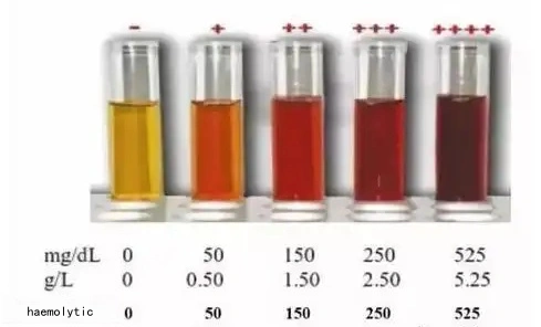
What Does Hemolysis Look Like?
Before diving into the science, let’s visualize what hemolysis looks like in practice.
| Hemolysis Level | Serum Appearance | Approximate Hemoglobin Concentration |
|---|---|---|
| None | Pale yellow (straw-colored) | <0.02 g/L |
| Mild | Slight pink tint | 0.02–0.05 g/L |
| Moderate | Reddish-pink | 0.05–0.3 g/L |
| Severe | Dark red to almost black | >0.3 g/L |
As hemolysis progresses, the serum color changes from pale yellow to deep red. Visually inspecting samples before use is the simplest way to identify potentially problematic specimens.
Why Does Hemolysis Occur?
Hemolysis can happen at various stages of blood collection and processing:
1. During Blood Collection
Using needles that are too small (increased shear stress)
Prolonged tourniquet application
Drawing blood from indwelling catheters
Vigorous mixing of collection tubes
2. During Processing
Delayed centrifugation (cells remain in contact with serum for too long)
Centrifugation at too high speed or incorrect temperature
Freezing whole blood instead of separated serum/plasma
3. Pathological Conditions
Certain diseases (e.g., hemolytic anemia, malaria)
In vivo hemolysis from mouse models with intraperitoneal bleeding
Serum vs. Plasma: Which Is More Prone to Hemolysis
Hemolysis is one of the most common pre-analytical errors that silently sabotages an ELISA test kit readout. When red blood cells rupture, they spill hemoglobin, proteases, and other intracellular molecules into the sample, creating a trio of problems: optical interference, enzymatic cross‑reactivity, and analyte degradation. If you are running a sensitive ELISA test kit — particularly one designed for low‑abundance biomarkers like C‑type natriuretic peptide (CNP) — even mild hemolysis can push your data from clear and publishable to uninterpretable.
Sometimes the test samples may be serum or plasma. For instance, our Human CNP ELISA Kit (Human C-Type Natriuretic Peptide Enzyme-Linked Immunosorbent Elisa Kit) and Rat C-Type Natriuretic Peptide (CNP) ELISA Kit can be used to quantitatively detect the concentration of CNP in human serum and plasma samples in vitro.The natural question that follows is: should I use serum or plasma to minimize the risk of hemolysis? The answer is not simply “both are fine if you have good technique.” In reality, under the same collection conditions, serum samples are significantly more susceptible to hemolysis than plasma samples. Understanding why gives you a practical lever to improve data quality.
Why Serum Is More Likely to Hemolyze
Serum preparation relies on the blood’s natural coagulation cascade, and this process is mechanically and chemically tough on red blood cells.
Clot retraction squeezes erythrocytes. As blood clots, activated platelets contract and pull the fibrin mesh tighter. This squeezing action exerts direct physical shear on fragile red cells trapped inside the clot, easily rupturing them. Plasma avoids this entirely because coagulation is inhibited from the start.
Longer processing window. Serum requires 30 minutes to 2 hours at room temperature for complete clotting. During this extended static period, temperature fluctuations, rough handling, or vibration can burst red cells. The longer blood sits without centrifugation, the greater the chance hemoglobin leaks into the supernatant.
Mistaken mixing. A surprisingly common error is treating a serum tube like an anticoagulant tube — inverting it repeatedly to “mix.” Without anticoagulant, this action grates red cells against irregular clot edges and can cause severe hemolysis in seconds.
Plasma: A Gentler Alternative, but Not Risk‑Free
Plasma is prepared by immediate centrifugation after mixing with an anticoagulant (EDTA, heparin, or citrate). Because clotting is blocked, the mechanical squeeze of clot retraction never happens, giving plasma an inherent advantage in preserving red cell integrity. However, plasma still demands care:
Delayed or incomplete mixing allows microclots to form, which can trap and damage red cells.
Chemical hemolysis can occur if anticoagulant concentrations are too high (e.g., potassium oxalate) or incompatible with the blood volume.
Lipemia or cold agglutinins may still cause interference, but these are independent of hemolysis.
How Hemolysis Directly Interferes with Your ELISA Test Kit
When a hemolyzed sample is loaded onto your ELISA test kit plate, three distinct mechanisms degrade the signal:
False‑positive background: Hemoglobin absorbs strongly at 450 nm, the detection wavelength of most TMB‑based ELISA kits. Even a faint pink tint can raise the optical density of negative wells, shrinking your signal‑to‑noise ratio. Worse, hemoglobin possesses pseudoperoxidase activity and can directly catalyze TMB substrate conversion, creating color that mimics a positive result.
HRP inhibition or competition: Some intracellular components released during hemolysis can chelate metal ions or inhibit horseradish peroxidase, partially or fully quenching the enzymatic reaction and producing falsely low signals.
Analyte degradation: Erythrocytes contain a rich cocktail of proteases. Once released into serum or plasma, these enzymes can cleave your target protein — particularly unstable peptides like CNP — leading to an under‑reported concentration that no ELISA test kit, however sensitive, can recover.
A Concrete Example: Human CNP ELISA
In our experience validating ELISA test kit products at Yanda Bio, hemolysis is especially detrimental when measuring CNP, a labile peptide with a short half‑life. A visibly hemolyzed serum sample can show a 30–50% reduction in measured CNP, not because the peptide was absent in vivo, but because it was degraded post‑collection. Simultaneously, the elevated background shortens the dynamic range, making it difficult to distinguish low‑level biological changes from noise.
Practical Recommendation
For most ELISA applications — and especially for CNP or similarly fragile biomarkers — we recommend plasma (EDTA or heparin) over serum. Plasma is faster to prepare (centrifuge within 15–30 minutes of collection), spares red cells from clot‑retraction trauma, and generally yields a cleaner matrix. If your protocol demands serum, allow clotting to proceed undisturbed at room temperature, centrifuge gently, and visually inspect for any pink discoloration. Never accept a hemolyzed sample for critical quantitative analysis.
Finally, always pair good sample handling with a robust ELISA test kit. Yanda Bio’s kits include detailed sample preparation guides and our team provides free technical consultation to help you adjust your pre‑analytical workflow. When you minimize hemolysis at the source, you get the accuracy, sensitivity, and reproducibility that your research deserves — and that our ELISA test kit manufacturing standards are built to deliver.
How Does Hemolysis Interfere with ELISA Results?
Hemoglobin, the primary contaminant in hemolyzed samples, can interfere with ELISA through two main mechanisms: false positives and false negatives.
Mechanism 1: False Positives—Hemoglobin Mimics HRP Activity
Most commercial ELISA kits, including mouse ELISA kits made in China, use horseradish peroxidase (HRP) as the enzyme conjugate. HRP catalyzes the conversion of the substrate 3,3′,5,5′-tetramethylbenzidine (TMB) from a colorless to a colored product.
The Chemistry Behind TMB Oxidation
Native TMB: Colorless, absorbance peak at 288 nm.
One-electron oxidation: TMB loses one electron, forming a blue charge-transfer complex (absorbance peaks at 370 nm and 652 nm).
Two-electron oxidation: After adding stop solution (acidification), TMB forms a yellow diimine product with a stable absorbance peak at 450 nm—the wavelength read in standard ELISA.
Hemoglobin has pseudo-peroxidase activity due to its heme group, which contains iron. This means hemoglobin can also catalyze TMB oxidation, mimicking the HRP reaction.
Data Point: A study comparing non‑hemolyzed and hemolyzed serum samples found that hemoglobin concentrations as low as 0.1 g/L increased background absorbance by 0.15–0.30 OD units at 450 nm, potentially pushing negative samples above the assay’s cutoff .
In a typical sandwich ELISA, this additional signal may be misinterpreted as specific antigen binding, leading to a false-positive result.
Mechanism 2: False Negatives—Interference with Antigen-Antibody Binding
Hemoglobin can also cause false negatives by physically blocking or competing for binding sites.
The “Musical Chairs” Analogy
Imagine the wells of your ELISA plate are chairs, and your target antigen is a person looking for a seat. Hemoglobin molecules, due to their high concentration and stickiness, can occupy these chairs non‑specifically. When the actual antigen arrives, there are no chairs left—so it remains unbound and is washed away.
In technical terms:
Hemoglobin can bind to the plate surface (blocking sites intended for capture antibodies)
Hemoglobin can interact with antibodies, reducing their effective concentration
Hemoglobin can form complexes with the target antigen, masking it from detection
Data Point: Research on cytokine measurements in hemolyzed samples showed that IL‑6 levels were underestimated by 20–40% in moderately hemolyzed samples compared to matched non‑hemolyzed controls . This underestimation can easily lead to false negatives, especially for low‑abundance analytes.
How to Prevent Sample Hemolysis
Prevention is always better than correction. Follow these best practices during blood collection and processing:
Method 1: Optimal Clotting and Centrifugation for Serum
Collect blood in tubes without anticoagulant (plain tubes or serum separator tubes).
Allow blood to clot at 37°C for 30 minutes in a constant‑temperature incubator.
Centrifuge at 5000 × g for 5–10 minutes. Adjust parameters based on your centrifuge model—larger centrifuges may require different speeds/times.
Carefully collect the supernatant (serum) without disturbing the pellet.
Method 2: Overnight Refrigeration
Place collected blood in a 4°C refrigerator overnight.
The next day, centrifuge and collect the supernatant.
This method yields clear serum with minimal hemolysis, especially useful for fragile samples.
Method 3: Use of Anticoagulants for Plasma
If you need plasma, choose appropriate anticoagulants:
Heparin (15–30 IU/mL) – good for most immunoassays
EDTA (1.5–2.2 mg/mL) – excellent for preserving cell morphology but may interfere with some metal‑dependent enzymes
Citrate (3.2%, 1:9 ratio) – specifically for coagulation studies
Always mix gently after collection (5–8 inversions) and centrifuge within 2 hours.
What If Hemolysis Has Already Occurred?
Despite best efforts, some samples may still be hemolyzed. Here are three approaches to salvage them:
Approach 1: Sample Dilution (Use with Caution)
Method: Dilute the hemolyzed sample with assay buffer before testing.
Pros: Simple, quick, and dilutes hemoglobin along with the target.
Cons: Also dilutes your analyte. If the target concentration is near the detection limit, dilution may push it below measurable levels.
Data Point: Diluting a moderately hemolyzed sample 1:2 reduces hemoglobin interference by ~50% but also reduces specific signal by ~50% . This “kill one thousand enemies at the cost of eight hundred of your own” approach should be used only when analyte concentrations are sufficiently high.
Approach 2: Add Hemolysis Inhibitors
Several commercial suppliers offer hemolysis interference inhibitors that block hemoglobin’s pseudo‑peroxidase activity.
Method: Pre‑treat samples with inhibitor according to manufacturer instructions before running the ELISA.
Pros: Specifically targets hemoglobin without diluting the analyte.
Cons: Adds an extra step; inhibitors may not be compatible with all assay formats.
Approach 3: Physical Removal of Hemoglobin
Method: Use centrifugation with ultrafiltration or dialysis to remove hemoglobin.
Ultrafiltration: Pass sample through a filter with a molecular weight cutoff (MWCO) smaller than hemoglobin (~64.5 kDa) but larger than your target protein.
Dialysis: Equilibrate sample against a large volume of buffer using a membrane that retains proteins above a certain size.
Pros: Can effectively remove hemoglobin.
Cons:
If your target protein is also >50 kDa, it may be retained along with hemoglobin.
Time‑consuming and may require optimization.
Also removes other small molecules (e.g., azide preservatives, if present).
When Hemolysis Is Unavoidable
Some experimental situations make hemolysis impossible to avoid. For example:
Ascites fluid from mice with intraperitoneal tumors often contains blood.
Mouse models of internal bleeding yield hemorrhagic samples.
Certain therapeutic agents cause red blood cell lysis in vivo.
In these cases, you must choose one of the salvage approaches above based on your specific analyte and experimental goals. Always include appropriate controls (e.g., spiked recovery experiments) to verify that your chosen method does not compromise data integrity.
Why Choose Yanda Bio Mouse ELISA Kits Made in China?
At Yanda Bio, we understand the challenges researchers face with real‑world samples—including hemolysis. That’s why our affodable mouse ELISA kits made in China are designed with robustness and reliability in mind.
Validated Performance: Each kit lot is tested against hemolyzed, lipemic, and icteric samples to ensure minimal interference. Our mouse IL‑6 elisa kit, for example, shows <15% change in OD with hemoglobin up to 0.2 g/L.
Comprehensive Menu: Over 6,000 detection targets covering immunology, oncology, neuroscience, metabolism, and infectious diseases. Whether you need to measure cytokines, chemokines, or niche biomarkers, we have the kit.
Custom Sensitivity: Struggling with low‑abundance targets? We can optimize detection limits—like our Mouse FDX1 (Adrenodoxin) ELISA Kit, now available with sensitivity down to 5 pg/mL.
Affordable Pricing: Standard kits from just $120. Bulk orders receive significant discounts.
Free Technical Support: Not sure how to handle hemolyzed samples? Our experienced scientists are just an email away.
Fast Delivery: Orders placed before 3:30 PM (China time) ship the same day; others ship next business day.
Conclusion
Hemolysis is a common but manageable challenge in ELISA experiments. By understanding its causes, recognizing its appearance, and knowing how it interferes with assay chemistry, you can take proactive steps to prevent or mitigate its effects.
Remember:
Prevent hemolysis through careful collection and processing.
Visually inspect samples before use.
Choose the right salvage method based on your sample and analyte.
Validate your approach with appropriate controls.
When you’re ready to run your assays, trust Yanda Bio mouse ELISA kits made in China for accurate, reproducible results—even with challenging samples.