
Key Features
- Superior Specificity for Equine IL-1β: No cross-reactivity or interference observed with equine IL-1β analogues. Highly specific for the target analyte.
- Wide Quantitative Range: 1.0 – 640 pg/mL, supported by a six-point standard curve (0, 40, 80, 160, 320, 640 pg/mL).
- High Sensitivity: Minimum detectable concentration < 1.0 pg/mL.
- Recombinant Protein Standards: Produced in a eukaryotic expression system, ensuring native-like conformation and optimal immunoreactivity.
- Validated Sample Types: Equine serum, EDTA/citrate/heparin plasma, cell culture supernatant, and tissue homogenates.
- Comprehensive Tissue Protocol: Includes mandatory protease inhibitor use, endogenous peroxidase inactivation (1% H₂O₂) for high-background tissues (e.g., liver, kidney), and low-temperature processing.
- Built-in 5× Sample Dilution: The assay automatically dilutes samples 5-fold (10 µL sample + 40 µL diluent). Multiply the interpolated result by 5 to obtain the actual concentration.
- One-Step Sandwich ELISA: Simultaneous incubation of sample/standard and HRP-conjugated detection antibody; total assay time approximately 2 hours.
- Reliable Performance: Intra- and inter-assay CV < 15%; standard curve linearity R ≥ 0.9900.
- Flexible Formats: 48-well and 96-well configurations with removable strips; up to 88 samples per plate (single wells).
- Storage & Stability: 6-month shelf life at 2–8°C, protected from light and moisture; cold-chain shipping.
Product Description
Interleukin-1β (IL-1β) is a pivotal pro-inflammatory cytokine that drives acute and chronic inflammation. In horses, IL-1β is a central mediator in osteoarthritis, laminitis, colitis, respiratory disease, and systemic inflammatory responses. Accurate measurement of equine IL-1β is essential for immunology research, drug development, and biomarker discovery.
The Yanda-HORSE-IL-1β ELISA Kit is a one-step double-antibody sandwich ELISA. A microplate pre-coated with an antibody specific for equine IL-1β captures the analyte from samples and standards. An HRP-labeled detection antibody is added simultaneously, and after washing, TMB substrate produces a colorimetric signal proportional to the IL-1β concentration. The absorbance is read at 450 nm, and sample concentrations are calculated from the standard curve.
Sample Collection & Preparation
Liquid Samples
| Sample Type | Procedure |
|---|---|
| Serum | Collect in pyrogen/endotoxin-free tubes. Let clot, then centrifuge at 3000 rpm for 10 min. Separate serum immediately. |
| Plasma | Use EDTA, citrate, or heparin. Centrifuge at 3000 rpm for 30 min; collect supernatant. |
| Cell Culture Supernatant | Centrifuge at 3000 rpm for 10 min to remove cells and debris. |
Tissue Homogenates – Standardized Protocol for Reliable Results
Critical: Protease Inhibitors Required
Tissues contain abundant proteases that rapidly degrade IL-1β. Freshly add a protease inhibitor cocktail (e.g., 1 mM PMSF) to the ice-cold homogenization buffer just before use.
Endogenous Peroxidase Inactivation
For tissues rich in endogenous peroxidases (e.g., liver, kidney, pancreas), high sample concentrations can react with the TMB substrate, causing false positives. After preparing the supernatant, treat it with 1% H₂O₂ for 15 minutes at room temperature to eliminate peroxidase activity.
Homogenization Steps:
- Rinse: Place fresh tissue on ice immediately; rinse with ice-cold PBS to remove residual blood.
- Homogenize: Add ice-cold lysis buffer (e.g., PBS or mild RIPA containing fresh protease inhibitors) at a ratio of 1 g tissue : 9 mL buffer. Homogenize thoroughly on ice. For complete lysis, sonicate or perform freeze-thaw cycles (keep samples ice-cold during sonication).
- Centrifuge: Centrifuge at 5000 × g for 5 minutes at 4°C. Carefully aspirate the clear middle supernatant for testing.
Storage
If not tested immediately, aliquot samples and store at -20°C. Avoid repeated freeze-thaw cycles. Thaw completely at room temperature and mix gently before use. Do not use samples containing sodium azide (NaN₃), as it inhibits HRP activity.
Critical Dilution Factor
The assay performs an automatic 5-fold dilution in the well (10 µL sample + 40 µL Sample Diluent). The concentration interpolated from the standard curve is the diluted value. Multiply the result by 5 to obtain the actual IL-1β concentration in the original sample.
The S0 standard (0 pg/mL) serves as the blank/negative control.
Baseline Reference Recommendation: We strongly advise performing a pilot experiment with a few known healthy equine serum/plasma samples (undiluted) to establish your laboratory’s own baseline reference range. If readings exceed the top standard (640 pg/mL after 5× correction), apply additional pre-dilution accordingly.
Technical Specifications
| Parameter | Specification |
|---|---|
| Standard Concentrations (S0–S5) | 0, 40, 80, 160, 320, 640 pg/mL |
| Quantitative Range | 1.0 – 640 pg/mL |
| Analytical Sensitivity | < 1.0 pg/mL |
| Accuracy (Linearity) | Standard curve R ≥ 0.9900 |
| Precision | Intra-assay CV < 15%; Inter-assay CV < 15% |
| Specificity | Highly specific for equine IL-1β. No cross-reactivity or interference with equine IL-1β analogues. |
| Sample Dilution Factor | 5× (automatically performed in the well) |
| Shelf Life | 6 months |
| Storage & Transport | 2–8°C, protected from light and moisture; cold-chain shipping |
Representative Standard Curve
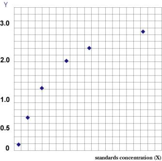
For spike-recovery, dilution linearity, and other validation data, please refer to the product instruction manual or contact technical support.
Kit Components
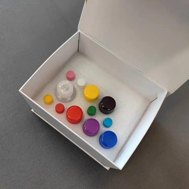
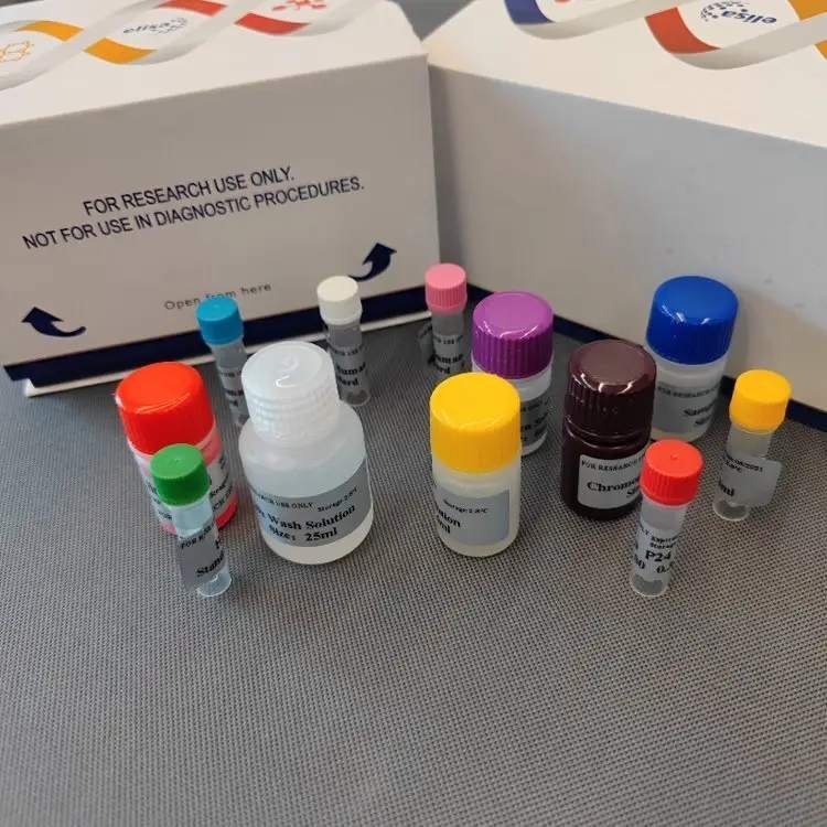
96-Well Format
| Component | Quantity | Notes |
|---|---|---|
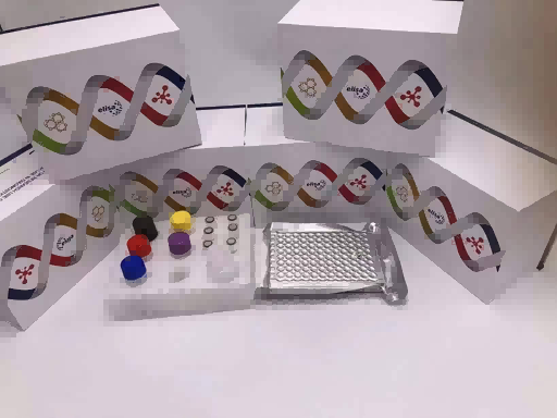
| Microelisa Stripplate | 12 strips × 8 wells | Pre-coated with anti-equine IL-1β antibody |
| Standard (0.3 mL/vial) | 6 vials | Ready-to-use liquid, 0–640 pg/mL |
| Sample Diluent | 6 mL | – |
| HRP-Conjugated Detection Antibody | 10 mL | Ready-to-use |
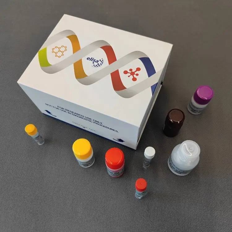
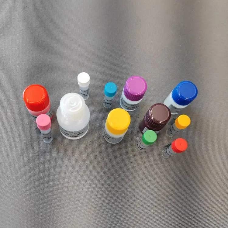
| 20× Wash Buffer | 25 mL | Dilute to 1× before use |
| Substrate A | 6 mL | – |
| Substrate B | 6 mL | – |
| Stop Solution | 6 mL | – |
| Plate Sealers | 2 sheets | – |
| Instruction Manual | 1 copy | – |
| Self-sealing Bag | 1 piece | For storing unused strips |
48-Well Format
| Component | Quantity | Notes |
|---|---|---|
| Microelisa Stripplate | 12 strips × 4 wells | Pre-coated, removable |
| Standard (0.3 mL/vial) | 6 vials | Ready-to-use liquid |
| Sample Diluent | 3 mL | – |
| HRP-Conjugated Detection Antibody | 5 mL | Ready-to-use |
| 20× Wash Buffer | 15 mL | Dilute to 1× |
| Substrate A | 3 mL | – |
| Substrate B | 3 mL | – |
| Stop Solution | 3 mL | – |
| Plate Sealers | 2 sheets | – |
| Instruction Manual | 1 copy | – |
| Self-sealing Bag | 1 piece | – |
Note: The 20× Wash Buffer may crystallize at 2–8°C; warm in a water bath until crystals dissolve before diluting to 1×.
Assay Procedure Summary
- Equilibrate: Let the foil pouch warm to room temperature for 20 min. Remove required strips; reseal unused strips and store at 4°C.
- Add Standards & Samples:
- Standard wells: 50 µL of each standard.
- Sample wells: 10 µL sample + 40 µL Sample Diluent.
- Blank well(s): Leave empty.
- Add Detection Antibody: Add 100 µL of HRP-conjugated detection antibody to all wells except blank. Cover with plate sealer and incubate at 37°C for 60 minutes.
- Wash: Aspirate and blot dry. Fill each well with 1× Wash Buffer, let stand for 1 min, then discard. Repeat for 5 washes (automated washer: 350 µL/well, 1-min soak).
- Substrate: Add 50 µL each of Substrate A and Substrate B. Incubate in the dark at 37°C for 15 minutes.
- Stop & Read: Add 50 µL Stop Solution. Read absorbance at 450 nm within 15 minutes.
Total hands-on time: approximately 2 hours.
Materials Required but Not Supplied:
- Microplate reader (450 nm)
- Adjustable pipettes and tips (0.5–1000 µL)
- 37°C incubator or water bath
- Distilled/deionized water
- Absorbent paper, wash bottle or automated plate washer
- Protease inhibitor cocktail (for tissue homogenates)
- 1% H₂O₂ (optional, for high-peroxidase tissues)
Throughput
| Format | Max. Number of Samples (Single Wells) |
|---|---|
| 48-well | 39–40 samples |
| 96-well | 85–88 samples |
Includes 6 standard wells and 1–2 blank wells. For duplicates, divide the sample count accordingly.
Expand Your Equine Cytokine Panel
This Horse IL-1β ELISA kit pairs effectively with other equine inflammatory markers. For multi‑cytokine profiling, consider these related products:
- Horse Interferon γ (IFN-γ) ELISA Kit – For the quantitative measurement of Horse Interferon γ (IFN-γ) levels in serum, plasma, cell culture supernatants, and tissue homogenates.
- Horse C-Reactive Protein (CRP) ELISA Kit – For the quantitative measurement of Horse C-Reactive Protein (CRP) levels in serum, plasma, cell culture supernatants, and tissue
- Horse Calcitonin (CT) ELISA Kit – Equine Calcitonin Quantitative Immunoassay for Calcium-Phosphorus Metabolism
You may also explore our full portfolio of equine ELISA kits covering growth factors, chemokines, and metabolic hormones.
Frequently Asked Questions
Q: Why must I multiply the result by 5?
A: The protocol dilutes the sample 5-fold directly in the well (10 µL sample + 40 µL diluent). The standard curve is constructed identically, so the interpolated value is the diluted concentration. Multiply by 5 to get the actual concentration.
Q: Is this kit specific for equine IL-1β? Will it detect IL-1α or other species?
A: The kit is highly specific for equine IL-1β. No cross-reactivity or interference has been observed with equine IL-1β analogues. It has been validated only for equine samples.
Q: How should I prepare tissue homogenates to avoid degradation or false positives?
A: Always add fresh protease inhibitors to your cold buffer and keep samples on ice. For liver, kidney, or pancreas, treat the supernatant with 1% H₂O₂ for 15 minutes to eliminate endogenous peroxidase activity. Centrifuge at 5000 × g at 4°C and use the clear supernatant.
Q: My sample reads above 640 pg/mL after multiplying by 5. What should I do?
A: Pre-dilute the sample further with the provided Sample Diluent (e.g., 1:2 or 1:5), re-assay, and multiply by the 5× in-well dilution and the additional dilution factor.
Q: Can I use hemolyzed or lipemic samples?
A: Hemolysis may interfere. We recommend collecting fresh samples without hemolysis. Lipemic samples should be centrifuged thoroughly; if turbid, recentrifuge before testing.
Ordering & Contact Information
| Item | Details |
|---|---|
| Catalog Number | Yanda-HORSE-IL-1β |
| Formats | 48-well / 96-well |
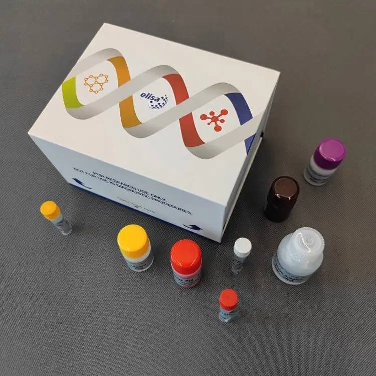
| Package Dimensions | 16 cm × 12.5 cm × 9.5 cm (neutral white box) |
| Shipping | Cold-chain transport (2–8°C), protected from light and moisture |
| Storage | 2–8°C; do not freeze |
| Shelf Life | 6 months |
| Intended Use | For research use only, not for diagnostic procedures |
Contact us
- WhatsApp: +86 198 3186 0669
- Email: songhawyble@gmail.com
For pricing, bulk orders, or customization inquiries, please reach out via the above channels.
Technical Documents & Support
- Instruction Manual – Download [insert link] or available upon request.
- MSDS – Supplied with the kit or available on request.
- Technical Support – For validation data, protocol optimization, or troubleshooting, contact us via WhatsApp or email.
Disclaimer
This product is for laboratory research use only and is not approved for clinical diagnosis, treatment, or prognosis in humans or animals. The manufacturer assumes no responsibility for consequences arising from improper use, protocol deviations, or unauthorized applications. Users are responsible for validating assay performance in their own sample matrices and experimental conditions.