
Important Pre-Analytical Notice: 2-AG Is a Lipophilic Endocannabinoid
2-Arachidonoylglycerol (2-AG) is a monoacylglycerol with extremely low aqueous solubility. Unlike standard protein or water-soluble lipid phosphate assays, direct homogenization in saline or PBS is not suitable for 2-AG extraction and will result in rapid degradation. This kit requires an organic solvent extraction step prior to the ELISA. Please read the sample preparation section carefully before use.
Key Features
- Direct Sandwich ELISA for 2-AG: Uses a pre-coated monoclonal antibody and HRP-detection antibody in a one-step sandwich format, optimized for the 2-AG hapten.
- Requires Organic Extraction: Lipids must be extracted from tissues or fluids using a solvent mixture (e.g., butanol:methanol:water or methanol/chloroform). Aqueous-only homogenization is not supported.
- High Sensitivity & Wide Range: Minimum detection < 0.1 nmol/L; quantitative range 0.1 – 40 nmol/L, with a six-point standard curve (0, 2.5, 5, 10, 20, 40 nmol/L).
- Built-in 5× Dilution in Well: 10 µL of reconstituted extract + 40 µL Sample Diluent; multiply the interpolated result by 5 to obtain the concentration in the assayed extract.
- Degradation Prevention Protocol: 2-AG is rapidly hydrolyzed by monoacylglycerol lipase (MAGL). The protocol mandates the addition of MAGL inhibitors (e.g., JZL184, MAFP) or broad-spectrum serine hydrolase inhibitors (PMSF) during sample collection, and strict low-temperature processing.
- Validated Sample Types: Mouse serum, plasma, cell culture supernatant, and tissue homogenates — with mandatory lipid extraction.
- Ready-to-Use Liquid Antibodies: Recombinant Antibodies supplied in liquid form; no reconstitution needed.
- Excellent Precision: Intra- and inter-assay CV < 15%; standard curve R ≥ 0.9900.
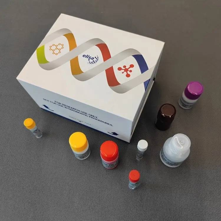
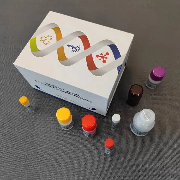
- Export-Ready: Neutral white box, cold-chain transport (2–8°C), 6-month shelf life; MSDS available.
Product Description
2-Arachidonoylglycerol (2-AG) is the most abundant endocannabinoid in the central nervous system and peripheral tissues. It acts as a full agonist at CB1 and CB2 cannabinoid receptors and modulates synaptic plasticity, pain, inflammation, and metabolism. Because 2-AG is rapidly isomerized to the biologically inactive 1-AG and hydrolyzed by MAGL, accurate quantitation requires both stabilization at collection and careful lipid extraction.
The Yanda-Mouse-2-AG ELISA Kit employs a one-step double-antibody sandwich ELISA with monoclonal antibodies specific for the 2-AG structure. The pre-coated plate captures 2-AG from reconstituted lipid extracts, and an HRP-labeled detection antibody is added simultaneously. After washing, TMB substrate produces a signal directly proportional to the 2-AG concentration, measured at 450 nm.
Sample Collection & Lipid Extraction
Critical Pre-Treatment to Preserve 2-AG
2-AG is highly unstable ex vivo. Follow these steps rigorously:
- Inhibitors: Immediately add MAGL inhibitor (e.g., JZL184, MAFP) or PMSF (1 mM) to the collection tube or homogenization solvent.
- Temperature: Perform all steps on ice. Freeze tissue in liquid nitrogen immediately after dissection.
- Avoid Aqueous Prolonged Contact: Do not homogenize directly in PBS or saline. Use organic solvents for extraction as described below.
Recommended Extraction Protocol
For tissues (e.g., brain):
- Weigh frozen tissue and add ice-cold organic solvent mixture: butanol:methanol:water (5:25:70, v/v/v) or methanol/chloroform (1:2, v/v). A common ratio is 1 mL solvent per 100 mg tissue.
- Homogenize on ice using a glass-Teflon homogenizer.
- Centrifuge at 3000–5000 × g for 10 min at 4°C to separate phases. Collect the organic phase (or the supernatant, depending on the mixture used) and evaporate if necessary, then reconstitute in the provided Sample Diluent or a compatible solvent as per the manual.
- Proceed to ELISA immediately or store reconstituted extracts at -20°C.
For plasma/serum:
- Add MAGL inhibitor to fresh blood, separate plasma at 4°C.
- Extract lipids using a similar solvent partitioning method (e.g., methanol/chloroform liquid-liquid extraction).
- Dry the organic layer under nitrogen and reconstitute in Sample Diluent.
Note: The exact reconstitution volume and compatibility should be tested in a pilot experiment. The final reconstituted sample must be free of solvents that could denature antibodies; the provided Sample Diluent is typically used for reconstitution.
Storage
Reconstituted extracts should be assayed immediately. If storage is necessary, keep at -20°C or -80°C in tightly sealed vials. Avoid repeated freeze-thaw.
Critical Dilution Factor
The assay includes a 5-fold dilution in the well: 10 µL of reconstituted sample + 40 µL Sample Diluent. The concentration read from the standard curve is the value in this diluted sample. Multiply the interpolated result by 5 to obtain the concentration in the reconstituted extract.
If you further diluted the extract before adding to the well, multiply by both the 5× in-well dilution and the additional factor.
Expected 2-AG Levels:
- Mouse brain: ~10–20 nmol/g tissue. After extraction and reconstitution, a high pre-dilution (e.g., 1:50 or 1:100) is usually needed to fall within the 0.1–40 nmol/L range.
- Mouse plasma: ~0.01–0.05 nmol/mL (10–50 pmol/mL). After extraction and reconstitution, less dilution may be required. Perform a pilot experiment to determine the optimal dilution.
Technical Specifications
| Parameter | Specification |
|---|---|
| Standard Concentrations (S0–S5) | 0, 2.5, 5, 10, 20, 40 nmol/L |
| Quantitative Range | 0.1 – 40 nmol/L |
| Analytical Sensitivity | < 0.1 nmol/L |
| Accuracy (Linearity) | Standard curve R ≥ 0.9900 |
| Precision | Intra-assay CV < 15%; Inter-assay CV < 15% |
| Specificity | Highly specific for 2-AG. No significant cross-reactivity or interference with structural analogues observed. |
| Sample Dilution Factor | 5× (in-well); additional pre-dilution often required |
| Shelf Life | 6 months |
| Storage & Transport | 2–8°C, protected from light and moisture; cold-chain shipping |
Representative Standard Curve
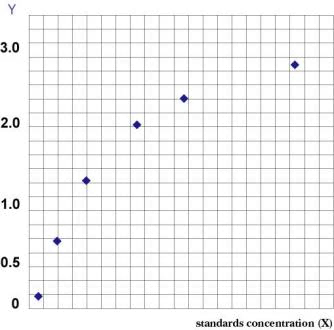
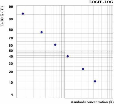
Detailed performance data are available in the product instruction manual. Contact technical support for further validation information.
Kit Components
96-Well Format
| Component | Quantity | Notes |
|---|---|---|
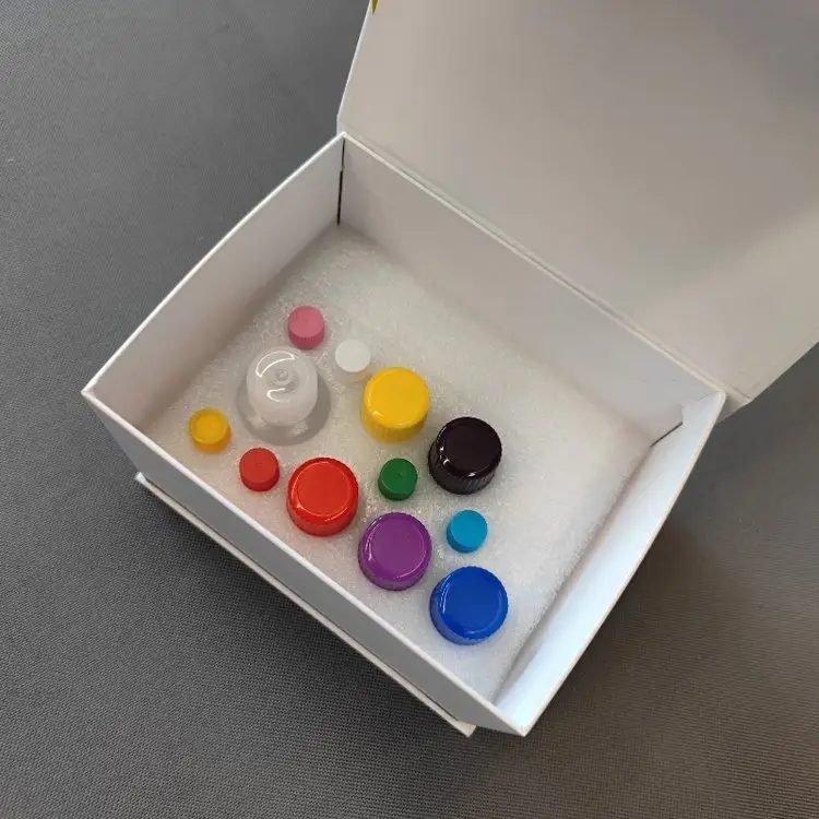
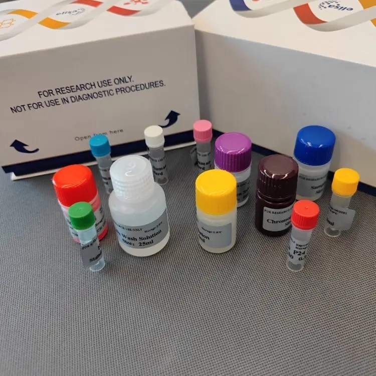
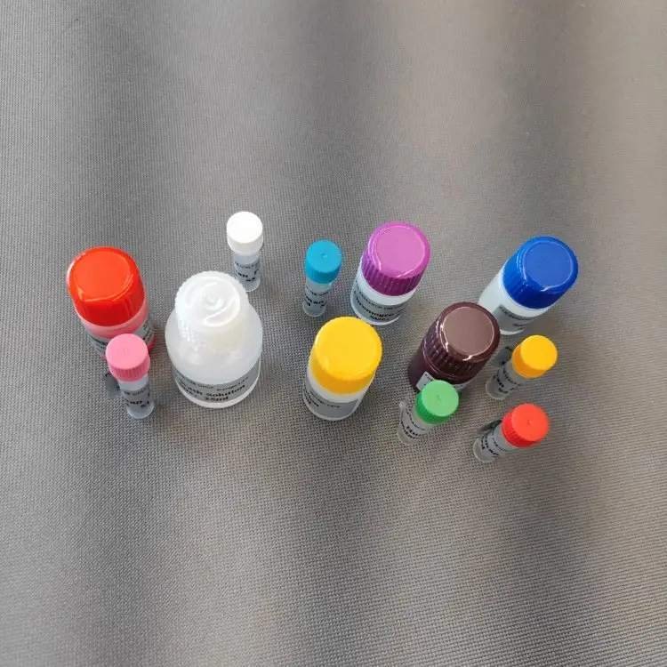
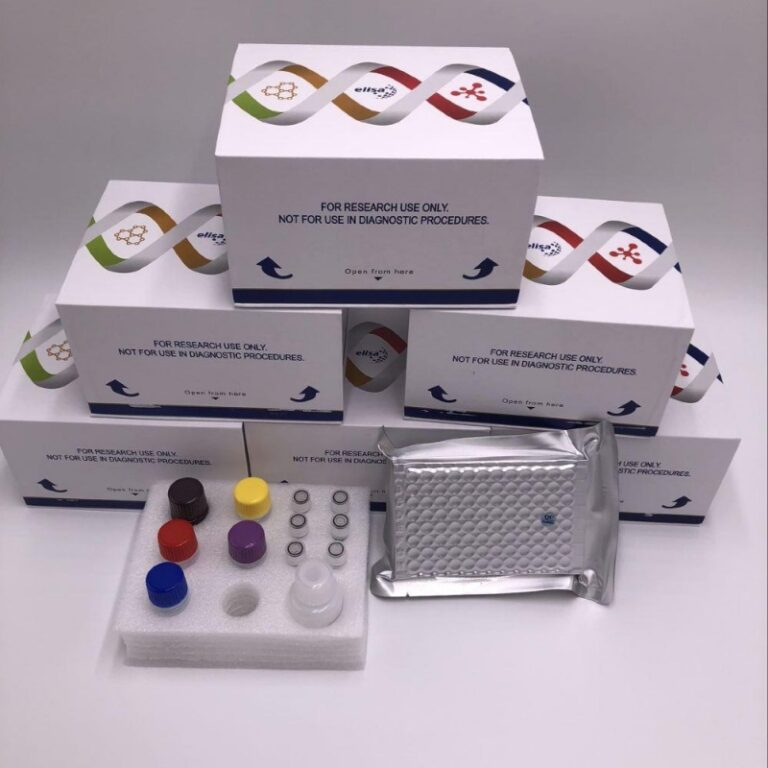
| Microelisa Stripplate | 12 strips × 8 wells | Pre-coated with anti-2-AG monoclonal antibody |
| Standard (0.3 mL/vial) | 6 vials | Ready-to-use liquid, 0–40 nmol/L |
| Sample Diluent | 6 mL | – |
| HRP-Conjugated Detection Antibody | 10 mL | Ready-to-use |
| 20× Wash Buffer | 25 mL | Dilute to 1× before use |
| Substrate A | 6 mL | – |
| Substrate B | 6 mL | – |
| Stop Solution | 6 mL | – |
| Plate Sealers | 2 sheets | – |
| Instruction Manual | 1 copy | – |
| Self-sealing Bag | 1 piece | For storing unused strips |
48-Well Format
| Component | Quantity | Notes |
|---|---|---|
| Microelisa Stripplate | 12 strips × 4 wells | Pre-coated |
| Standard (0.3 mL/vial) | 6 vials | Ready-to-use liquid |
| Sample Diluent | 3 mL | – |
| HRP-Conjugated Detection Antibody | 5 mL | Ready-to-use |
| 20× Wash Buffer | 15 mL | Dilute to 1× |
| Substrate A | 3 mL | – |
| Substrate B | 3 mL | – |
| Stop Solution | 3 mL | – |
| Plate Sealers | 2 sheets | – |
| Instruction Manual | 1 copy | – |
| Self-sealing Bag | 1 piece | – |
Crystallization of 20× Wash Buffer upon cold storage is normal; warm in a water bath until crystals dissolve before dilution.
Assay Procedure Summary
- Reconstitute lipid extract in an appropriate volume of Sample Diluent (or as per manual).
- Equilibrate: Warm the foil pouch to room temperature for 20 min. Remove required strips; reseal unused strips at 4°C.
- Add Standards & Samples:
- Standard wells: 50 µL of each standard.
- Sample wells: 10 µL of reconstituted sample + 40 µL Sample Diluent.
- Blank well(s): Leave empty.
- Add Detection Antibody: Dispense 100 µL HRP-conjugated detection antibody into all wells except blank. Seal and incubate at 37°C for 60 minutes.
- Wash: Aspirate, blot dry. Fill wells with 1× Wash Buffer, soak 1 min, discard. Repeat 5 times (automated: 350 µL/well).
- Substrate: Add 50 µL each of Substrate A and Substrate B. Incubate in the dark at 37°C for 15 minutes.
- Stop & Read: Add 50 µL Stop Solution. Measure absorbance at 450 nm within 15 min.
Total hands-on time: ~2 hours (excluding lipid extraction).
Materials Required but Not Supplied:
- Microplate reader (450 nm)
- Pipettes (0.5–1000 µL)
- 37°C incubator or water bath
- Distilled/deionized water
- Organic solvents (butanol, methanol, chloroform), nitrogen evaporator (optional)
- MAGL inhibitor (JZL184, MAFP) or PMSF
- Homogenizer, centrifuge, glass vials
Throughput
| Format | Max. Samples (Single Wells) |
|---|---|
| 48-well | 39–40 |
| 96-well | 85–88 |
Includes 6 standard wells and 1–2 blank wells. For duplicates, divide accordingly.
Related Mouse ELISA Kit:
- Mouse Osteopontin (SPP1) ELISA Kit —— Quantify a Key Inflammatory & Bone Marker
- Mouse 1,25-Dihydroxyvitamin D3 ELISA Kit
- Mouse Ribonucleoprotein RNP ELISA Kit – Essential Tool for Autoimmune & Molecular Biology Research
- Mouse RAS domain-containing protein 6 (RASSF6) ELISA Kit – For Cancer & Cellular Signaling Research
Frequently Asked Questions
Q: Can I homogenize brain tissue in PBS and measure directly?
A: No. 2-AG is lipophilic and will not be efficiently extracted by aqueous buffers. You must perform lipid extraction using an organic solvent mixture (e.g., butanol:methanol:water or methanol/chloroform) as described in the manual.
Q: How do I prevent 2-AG degradation during sample collection?
A: Add MAGL inhibitors (JZL184, MAFP) or PMSF immediately. Keep samples on ice or freeze in liquid nitrogen. Process promptly and avoid prolonged exposure to water.
Q: Does this kit cross-react with 1-AG or anandamide?
A: No significant cross-reactivity with structural analogues has been observed in validation studies. For detailed cross-reactivity data, please contact technical support.
Q: What are the expected 2-AG levels in mouse brain and plasma?
A: Brain 2-AG levels are typically 10–20 nmol/g tissue; plasma levels are much lower, around 0.01–0.05 nmol/mL. Brain extracts will require high pre-dilution (e.g., 1:50–1:100) to fit the standard curve.
Q: Can I use this kit for rat or human samples?
A: This kit is validated for mouse samples. Cross-reactivity with other species has not been established. Please inquire about species-specific options.
Q: Do you provide MSDS and composition documents for export?
A: Yes, these documents are available upon request. The kit is shipped in a neutral white box with cold-chain packaging.
Ordering & Contact Information
| Item | Details |
|---|---|
| Catalog Number | Yanda-Mouse-2-AG |
| Formats | 48-well / 96-well |
| Package Dimensions | 16 cm × 12.5 cm × 9.5 cm (neutral white box) |
| Shipping | Cold-chain (2–8°C), protected from light and moisture |
| Storage | 2–8°C; do not freeze |
| Shelf Life | 6 months |
| Intended Use | For research use only, not for diagnostic procedures |
Contact us
- WhatsApp: +86 198 3186 0669
- Email: songhawyble@gmail.com
For pricing, bulk orders, or species-specific kits, please reach out directly.
Technical Documents & Support
Instruction Manual
- MSDS & Certificate of Composition – Available upon request.
- Technical Support – For extraction protocols, inhibitor recommendations, or data analysis, contact us via WhatsApp or email.
Disclaimer
This product is for laboratory research use only and is not approved for clinical diagnosis, treatment, or prognosis. The manufacturer assumes no liability for improper use, protocol deviations, or unauthorized applications. Users are responsible for validating assay performance in their own sample matrices and experimental conditions.