
Key Features
- Total Rabbit IgG Detection: Measures total immunoglobulin G without distinguishing subclasses (IgG1, IgG2, etc.), making it suitable for broad humoral immunity studies.
- High Specificity, Minimal Interference: No significant cross-reactivity or interference with rabbit IgM or IgA. Designed for unambiguous IgG quantitation in complex matrices.
- Optimized for High-Abundance Samples: Rabbit serum contains exceptionally high IgG levels (5–20 mg/mL). A dedicated pre-dilution strategy (typically 1:1,000–1:10,000) is provided to bring readings into the standard curve.
- Wide g/L Scale: Standard curve from 0 to 24 g/L (6 points), quantitative range 0.1–24 g/L, sensitivity <0.1 g/L.
- Recombinant Rabbit IgG Standard: Produced in a eukaryotic system for native conformation and reliable immunoreactivity.
- Built-in 5× Dilution: In-well dilution (10 µL sample + 40 µL diluent) simplifies workflow; multiply the interpolated concentration by 5 and any additional pre-dilution factor to obtain actual values.
- Validated Sample Types: Rabbit serum, EDTA/citrate/heparin plasma, cell culture supernatant, and tissue homogenates.
- Special Tissue Handling: Organ perfusion with PBS is strongly advised to remove blood-derived IgG; guidelines for homogenization and avoidance of NaN₃ preservatives are included.
- One-Step Sandwich ELISA: Simultaneous incubation of sample/standard and HRP-conjugated detection antibody; total assay time ~2 hours.
- Reproducible Performance: Intra- and inter-assay CV <15%; standard curve linearity R ≥0.9900.
- Flexible Throughput: 48-well and 96-well removable-strip plates; up to 88 samples per plate (single wells).
- Shelf Life: 6 months at 2–8°C, protected from light and moisture; cold-chain shipping.
Product Description
Immunoglobulin G (IgG) is the most abundant antibody isotype in rabbit serum and a cornerstone of humoral immunity. It is widely measured in vaccine development, monoclonal/polyclonal antibody production, inflammation models, immune status and diagnostic reagent research. Accurate total IgG quantitation in rabbits requires an assay that handles exceptionally high concentrations without sacrificing specificity.
The Yanda-RABBIT-IgG ELISA Kit utilizes a one-step double-antibody sandwich ELISA. A capture antibody specific for rabbit IgG is pre-coated on the plate. Samples and standards are co-incubated with an HRP-labeled detection antibody. After washing, TMB substrate generates a colorimetric signal proportional to IgG concentration, measured at 450 nm.
Note: This kit detects total rabbit IgG and does not differentiate between IgG subclasses. It has been validated exclusively for rabbit samples; cross-species use is not recommended.
Sample Collection & Preparation
General Guidelines
| Sample Type | Procedure |
|---|---|
| Serum | Collect blood in pyrogen/endotoxin-free tubes without anticoagulant. Allow to clot, centrifuge at 3000 rpm for 10 min. Separate serum immediately. |
| Plasma | Use EDTA, citrate, or heparin. Centrifuge at 3000 rpm for 30 min; collect supernatant. |
| Cell Culture Supernatant | Centrifuge at 3000 rpm for 10 min to remove cells and debris. |
| Tissue Homogenate | Perfuse organ with ice-cold PBS to remove blood. Mince, add PBS or lysis buffer (1:9 w/v), homogenize on ice, centrifuge at 3000 rpm for 10 min at 4°C. Collect supernatant. |
Critical Pre-Dilution for Rabbit Serum & Plasma
Rabbit IgG levels in serum/plasma typically fall between 5 and 20 mg/mL, far exceeding the assay’s 24 g/L top standard (after 5× in-well dilution, which translates to ~120 g/L for the original sample). Direct testing without additional pre-dilution will saturate the standard curve.
Recommended pre-dilution: Perform a pilot experiment by serially diluting serum/plasma 1:1,000 to 1:10,000 (or higher) in Sample Diluent before adding to the well. Choose the dilution that yields an OD in the middle of the standard curve. Multiply the final calculated concentration by the pre-dilution factor and the 5× in-well dilution.
For cell supernatant and tissue homogenates, IgG levels may be lower; start with a pilot dilution series (e.g., neat, 1:2, 1:4, 1:10) to determine the optimal factor.
Interfering Substances & Quality Control
- NaN₃ Prohibition: Sodium azide at concentrations >0.1% inhibits HRP. Do not use NaN₃ as a preservative in samples.
- Hemolysis & Lipemia: Hemolysis or grossly lipemic samples cause light scattering and interfere with 450 nm absorbance. Centrifuge such samples at 3000 rpm for 10 minutes to remove cell debris and chylomicrons before testing. Interpret results with caution.
- Storage: Aliquot and store at -20°C. Avoid repeated freeze-thaw cycles. Thaw completely at room temperature and mix gently prior to use.
Dilution & Calculation Guide
- In-well dilution: 10 µL sample + 40 µL Sample Diluent = 5× dilution automatically.
- Additional pre-dilution: If you pre-diluted the sample, for example, 1:2,000 before adding to the well, the total dilution factor is 5 × 2,000 = 10,000.
- Final concentration = Interpolated value from standard curve (in g/L) × 5 × pre-dilution factor.
Example: A rabbit serum sample pre-diluted 1:5,000 gives a reading of 0.6 g/L after 5× in-well dilution.
Final IgG concentration = 0.6 × 5 × 5,000 = 15,000 g/L? Wait, that seems off. Let’s recalculate: Actually, the interpolated concentration is already the concentration in the diluted sample (after 5× dilution). If further pre-dilution of 1:5,000 was applied, the original concentration = (interpolated value in g/L) × 5 × 5,000. But 0.6 g/L × 5,000 = 3,000 g/L, then ×5? No, let’s think: The assay’s standard curve corresponds to the well contents. The well contains 10 µL of the pre-diluted sample plus 40 µL diluent, so the concentration in that well represents the concentration of the pre-diluted sample. So if pre-dilution is 1:5,000, the concentration in the pre-diluted sample is what the curve reads. To get back to original: multiply by the pre-dilution factor (5,000). The ×5 is already accounted for if the standard curve is constructed with the same in-well dilution. The standard curve is built with 50 µL of standard, while sample wells have 10 µL sample + 40 µL diluent. The standard curve is not diluted that way; the standard is added neat. So the dilution factor for sample vs standard is 5×. Therefore, the interpolated concentration from the standard curve must be multiplied by 5 to get the concentration in the pre-diluted sample. Then multiply by the pre-dilution factor. So total factor = 5 × pre-dilution factor. For 1:5,000 pre-dilution, total factor = 25,000. 0.6 g/L × 25,000 = 15,000 g/L, which is 15 g/mL. That matches rabbit serum levels. Good. So the guide is correct: multiply by 5 and by any extra pre-dilution factor.
I’ll include a clear formula in the page.
Technical Specifications
| Parameter | Specification |
|---|---|
| Standard Concentrations (S0–S5) | 0, 1.5, 3, 6, 12, 24 g/L |
| Quantitative Range | 0.1 – 24 g/L |
| Analytical Sensitivity | < 0.1 g/L |
| Accuracy (Linearity) | Standard curve R ≥ 0.9900 |
| Precision | Intra-assay CV < 15%; Inter-assay CV < 15% |
| Specificity | Highly specific for rabbit total IgG. No significant cross-reactivity or interference with rabbit IgM or IgA. Not tested for IgG subclass discrimination. |
| Species Restriction | Validated only for rabbit samples; not recommended for other species. |
| Sample Dilution Factor | 5× (in-well); additional pre-dilution typically required for serum/plasma |
| Shelf Life | 6 months |
| Storage & Transport | 2–8°C, protected from light and moisture; cold-chain shipping |
Representative Standard Curve
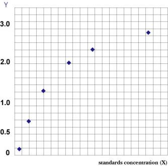
Representative validation data are available in the product instruction manual. Contact technical support for detailed performance data.
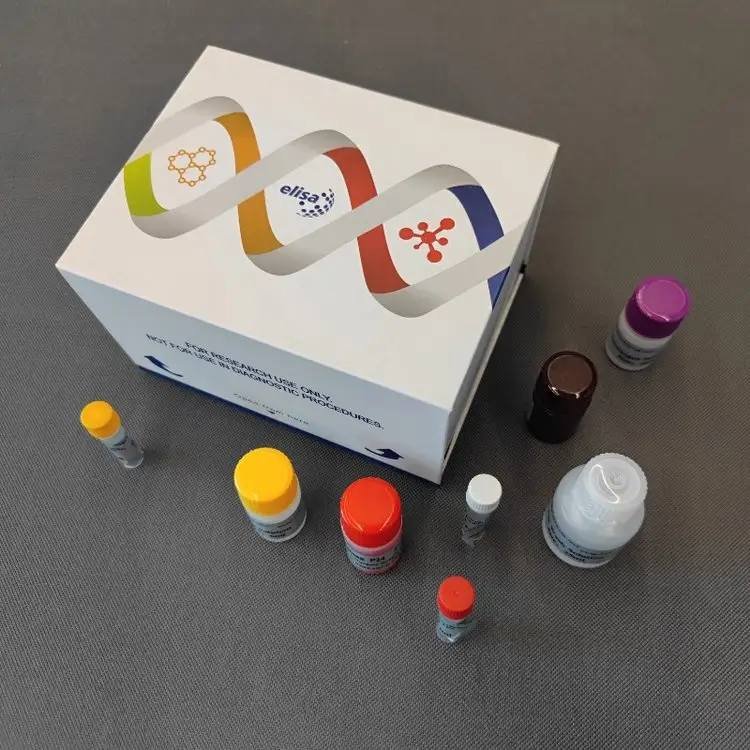
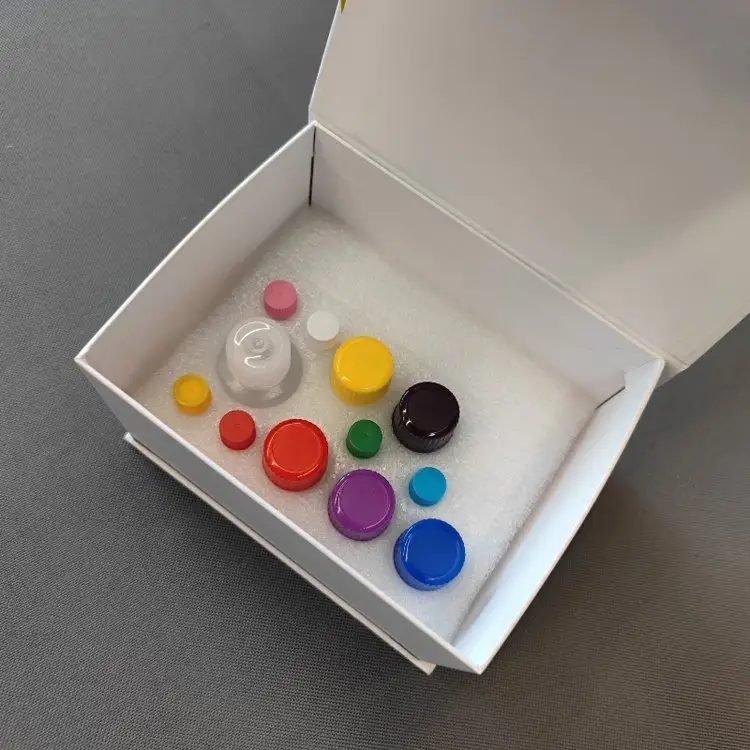
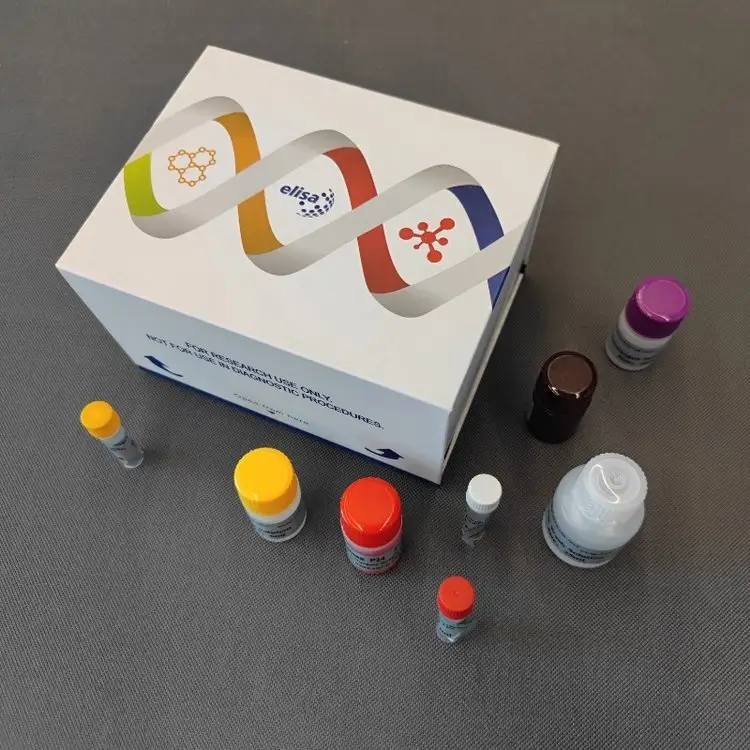
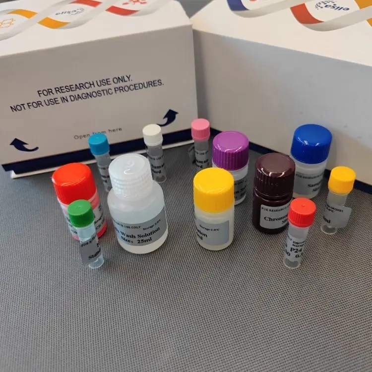
Kit Components
96-Well Format
| Component | Quantity | Notes |
|---|---|---|
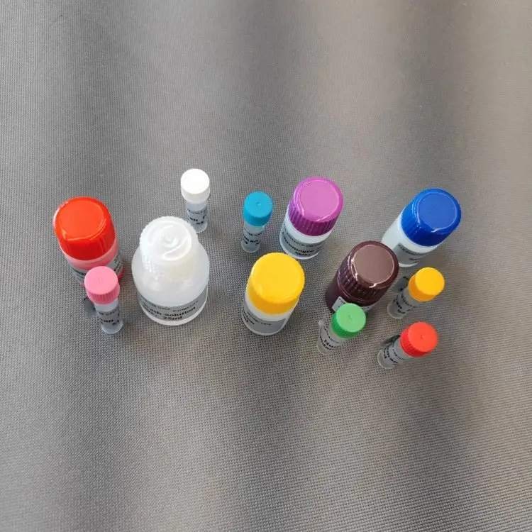
| Microelisa Stripplate | 12 strips × 8 wells | Pre-coated with anti-rabbit IgG antibody |
| Standard (0.3 mL/vial) | 6 vials | Ready-to-use liquid, 0–24 g/L |
| Sample Diluent | 6 mL | – |
| HRP-Conjugated Detection Antibody | 10 mL | Ready-to-use |
| 20× Wash Buffer | 25 mL | Dilute to 1× before use |
| Substrate A | 6 mL | – |
| Substrate B | 6 mL | – |
| Stop Solution | 6 mL | – |
| Plate Sealers | 2 sheets | – |
| Instruction Manual | 1 copy | – |
| Self-sealing Bag | 1 piece | For unused strip storage |
48-Well Format
| Component | Quantity | Notes |
|---|---|---|
| Microelisa Stripplate | 12 strips × 4 wells | Pre-coated, removable |
| Standard (0.3 mL/vial) | 6 vials | Ready-to-use liquid |
| Sample Diluent | 3 mL | – |
| HRP-Conjugated Detection Antibody | 5 mL | Ready-to-use |
| 20× Wash Buffer | 15 mL | Dilute to 1× |
| Substrate A | 3 mL | – |
| Substrate B | 3 mL | – |
| Stop Solution | 3 mL | – |
| Plate Sealers | 2 sheets | – |
| Instruction Manual | 1 copy | – |
| Self-sealing Bag | 1 piece | – |
Crystallization of 20× Wash Buffer is normal; warm in a water bath to dissolve.
Assay Procedure Summary
- Equilibrate: Bring the foil pouch to room temperature for 20 min. Remove required strips; reseal unused strips at 4°C.
- Add Standards & Samples:
- Standard wells: 50 µL of each standard.
- Sample wells: 10 µL of pre-diluted sample + 40 µL Sample Diluent.
- Blank well(s): leave empty.
- Add Detection Antibody: Add 100 µL HRP-conjugated detection antibody to all wells except blank. Seal and incubate at 37°C for 60 minutes.
- Wash: Aspirate, blot dry. Fill wells with 1× Wash Buffer, soak 1 min, discard. Repeat 5 times (automated: 350 µL/well, 1-min soak).
- Substrate: Add 50 µL Substrate A and 50 µL Substrate B. Incubate in the dark at 37°C for 15 minutes.
- Stop & Read: Add 50 µL Stop Solution. Read absorbance at 450 nm within 15 min.
Total assay time: ~2 hours.
Materials Required but Not Supplied:
- Microplate reader (450 nm)
- Adjustable pipettes (0.5–1000 µL) and tips
- 37°C incubator or water bath
- Distilled/deionized water
- Absorbent paper, wash bottle or automated washer
- Protease inhibitors (optional, for tissue)
Throughput
| Format | Max. Samples (Single Wells) |
|---|---|
| 48-well | 39–40 |
| 96-well | 85–88 |
Includes 6 standard and 1–2 blank wells. For duplicates, halve the sample count.
Discover More ELISA Kits:
- Rabbit ELISA Kits Collection — Browse our complete rabbit product line
- Rabbit Immunoglobulin A (IgA) ELISA Kit — For mucosal antibody studies
- Rabbit Immunoglobulin M (IgM) ELISA Kit — For primary immune response detection
- Human Immunoglobulin G (IgG) ELISA Kit – High‑Precision Sandwich Assay for Humoral Immunity Assessment
Frequently Asked Questions
Q: Why do I need to pre-dilute my rabbit serum before the assay?
A: Rabbit IgG concentrations (5–20 mg/mL) are extremely high. The built-in 5× dilution alone is insufficient to bring levels into the 0–24 g/L range. We recommend a pre-dilution of 1:1,000 to 1:10,000 or higher, determined by pilot experiment.
Q: How do I calculate the final IgG concentration?
A: Final concentration (g/L) = [interpolated value from standard curve] × 5 (in-well dilution) × additional pre-dilution factor. For example, if you pre-diluted 1:5,000 and the interpolated value is 0.6 g/L, the final concentration = 0.6 × 5 × 5,000 = 15,000 g/L (15 mg/mL). If no extra pre-dilution was performed, just multiply by 5.
Q: Can this kit distinguish between rabbit IgG subclasses?
A: No. It measures total rabbit IgG. Subclass-specific reactivity (IgG1, IgG2, etc.) has not been validated.
Q: Does it cross-react with rabbit IgM or IgA?
A: No significant cross-reactivity or interference has been observed with rabbit IgM or IgA.
Q: Can I use this kit for mouse or human IgG?
A: No. It is validated only for rabbit samples. Cross-reactivity with other species has not been tested and is not expected to be reliable.
Q: How should I handle hemolyzed or lipemic samples?
A: These can interfere with OD readings. Centrifuge at 3000 rpm for 10 min to remove particulates before testing. Avoid using severely hemolyzed or turbid samples.
Q: Why is NaN₃ prohibited?
A: Sodium azide (>0.1%) inhibits HRP activity, causing falsely low signals. Do not use it as a preservative.
Q: For tissue samples, why is PBS perfusion recommended?
A: Rabbit tissues contain blood rich in IgG. Without perfusion, blood-derived IgG will obscure the endogenous tissue signal. Perfuse organs with cold PBS before homogenization.
Ordering & Contact
| Item | Details |
|---|---|
| Catalog Number | Yanda-RABBIT-IgG |
| Formats | 48-well / 96-well |
| Package Dimensions | 16 cm × 12.5 cm × 9.5 cm (neutral white box) |
| Shipping | Cold-chain (2–8°C), protected from light and moisture |
| Storage | 2–8°C; do not freeze |
| Shelf Life | 6 months |
| Intended Use | For research use only, not for diagnostic procedures |
Contact us
- WhatsApp: +86 198 3186 0669
- Email: songhawyble@gmail.com
For pricing, bulk orders, or customization, please reach out directly.
Technical Documents & Support
- MSDS – Supplied with the kit or upon request.
- Technical Support – For protocol optimization or troubleshooting, contact us on WhatsApp or via email.
Disclaimer
This product is for laboratory research use only and is not approved for clinical diagnosis, treatment, or prognosis. The ELISA kit manufacturer assumes no liability for improper use, protocol deviations, or unauthorized applications. Users are responsible for validating assay performance in their own sample matrix and experimental conditions.