
Assay Principle — Competitive ELISA for a Transient Second Messenger
Inositol 1,4,5-trisphosphate (IP3) is a small-molecule second messenger (~420 Da) that is too small for traditional sandwich ELISA. This kit utilizes a competitive inhibition enzyme immunoassay. The microplate is pre-coated with an anti-IP3 antibody. Free IP3 in the sample competes with an HRP-labeled IP3 conjugate for a limited number of antibody binding sites. The resulting OD signal is inversely proportional to the IP3 concentration in the sample.
Note on standard concentrations: The standard curve values (0–480 pg/mL) refer to in-well final concentrations. Actual sample concentrations must be calculated by multiplying the interpolated value by the appropriate dilution factor.
Key Features
- Detects IP3 via Competitive ELISA: Overcomes the size limitation of IP3 that prevents sandwich detection. High specificity is achieved through antibody competition rather than dual-antibody binding.
- No Cross‑Reactivity with Related Inositol Phosphates: No significant interference or cross‑reactivity with IP2, IP4, PIP2, or other inositol phosphate analogues.
- Comprehensive Degradation Prevention Protocol: IP3 is rapidly hydrolyzed by endogenous phosphatases. The kit provides explicit guidance on phosphatase inhibitors (NaF 10–20 mM, sodium orthovanadate 1 mM, or commercial cocktail) and rapid sample processing on ice.
- Acid Extraction Compatible: For maximum IP3 stabilization, samples can be extracted with perchloric acid (PCA) or trichloroacetic acid (TCA) and neutralized to pH ~7.4 before assay. PBS‑based homogenization with inhibitors is also supported as a standard method.
- Cell Stimulation Sampling Guide: IP3 peaks within seconds of GPCR activation. Recommended sampling time points: 30 sec, 1 min, 5 min. Immediately quench with ice‑cold extraction buffer containing phosphatase inhibitors.
- Ready‑to‑Use Liquid Standards: Six standards supplied (0, 30, 60, 120, 240, 480 pg/mL); no reconstitution required.
- High Sensitivity & Defined Range: Quantitative range 1.0–480 pg/mL (in‑well), sensitivity <1.0 pg/mL.
- Reliable Precision: Intra‑ and inter‑assay CV <15%; standard curve R ≥0.9900.
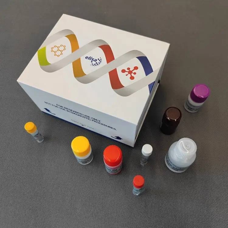
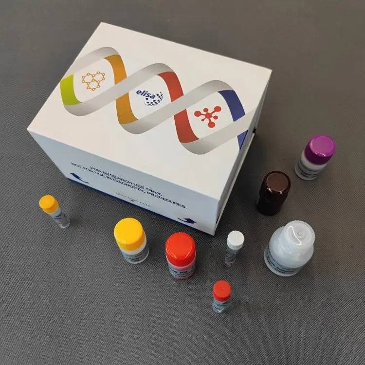
- Export‑Ready: Neutral white box (16 × 12.5 × 9.5 cm), cold‑chain shipping (2–8°C), 6‑month shelf life. MSDS available upon request.
Product Description
IP3 is a critical second messenger generated by phospholipase C (PLC)‑mediated hydrolysis of phosphatidylinositol 4,5‑bisphosphate (PIP2). Upon binding to IP3 receptors on the endoplasmic reticulum, IP3 triggers the release of Ca²⁺ into the cytoplasm, initiating a cascade of cellular responses including contraction, secretion, metabolism, and gene expression. In human research, IP3 measurement is essential for studying GPCR signaling, calcium dynamics, and phosphoinositide metabolism.
The Yanda-Human-IP3 ELISA Kit provides a convenient competitive ELISA format to quantify IP3 in cell lysates and tissue extracts without the need for LC‑MS/MS instrumentation. It is intended exclusively for research applications.Please select the appropriate cell lysis solution.
Sample Collection & Preparation
Recommended Sample Types
- Stimulated cell lysates (strongly recommended) — IP3 is an intracellular signaling molecule; levels in resting cells are low and spike rapidly after stimulation.
- Tissue homogenates — Process immediately after dissection.
- Serum / plasma — Not recommended. Circulating IP3 is unstable and present at extremely low concentrations; reliable quantitation is difficult.
Phosphatase Inhibitor Protocol (Mandatory for IP3 Stability)
IP3 is rapidly degraded in vitro. Add fresh phosphatase inhibitors to all extraction buffers:
| Inhibitor | Recommended Working Concentration |
|---|---|
| Sodium fluoride (NaF) | 10–20 mM |
| Sodium orthovanadate (Na₃VO₄) | 1 mM |
| Phosphatase Inhibitor Cocktail (100×) | Use at 1× final concentration (contains NaF, orthovanadate, sodium pyrophosphate, β‑glycerophosphate, etc.) |
Standard PBS‑Based Extraction
- Wash cells with ice‑cold PBS. For tissue, mince and rinse with ice‑cold PBS.
- Add ice‑cold PBS containing fresh phosphatase inhibitors (± protease inhibitors).
- Lyse by repeated freeze‑thaw cycles or sonication on ice.
- Centrifuge at 3000 rpm for 10 min at 4°C. Collect supernatant for immediate assay or freeze at -20°C.
Acid Extraction (Enhanced Stability)
For maximum phosphatase inactivation, PCA or TCA extraction can be used:
- Add ice‑cold 1.5 M PCA (or 10% TCA) to cell pellet or tissue, vortex.
- Incubate on ice for 15 min, then centrifuge at 12,000 × g for 10 min at 4°C.
- Neutralize the supernatant with 2 M KOH (or 1 M Tris base) until pH ~7.4. Check with pH paper.
- Remove precipitated potassium perchlorate by centrifugation if necessary. Use the neutralized supernatant for ELISA.
Cell Stimulation Time Window
For GPCR‑mediated IP3 generation:
- Collect samples at 30 sec, 1 min, and 5 min after agonist addition.
- Immediately aspirate medium and add ice‑cold extraction buffer (PBS + inhibitors or acid) directly to the culture dish/plate.
- Keep all subsequent steps at 4°C.
Storage & NaN₃ Prohibition
- Aliquot and store at -20°C. Avoid repeated freeze‑thaw cycles.
- Do not use sodium azide (NaN₃) — it is a potent HRP inhibitor and will cause false‑negative results.
Dilution & Calculation
- In‑well dilution: 10 μL sample + 40 μL Sample Diluent = 5‑fold dilution.
- Standard curve: Values (0–480 pg/mL) are in‑well concentrations. The interpolated value from the standard curve is the IP3 concentration in the well.
- Final sample concentration = In‑well interpolated value × 5 (in‑well dilution) × additional pre‑dilution factor.
If acid‑extracted and neutralized samples were further diluted before loading, multiply by that factor as well.
Data Fitting: Because the curve is inverse, use 4‑parameter logistic (4PL) or log‑logit fitting (e.g., GraphPad Prism, MyCurveFit).
Technical Specifications
| Parameter | Specification |
|---|---|
| Standard Concentrations (S0–S5) | 0, 30, 60, 120, 240, 480 pg/mL (in‑well) |
| Quantitative Range | 1.0 – 480 pg/mL (in‑well) |
| Analytical Sensitivity | < 1.0 pg/mL (in‑well) |
| Accuracy (Linearity) | Standard curve R ≥ 0.9900 |
| Precision | Intra‑assay CV < 15%; Inter‑assay CV < 15% |
| Specificity | Highly specific for human IP3. No significant cross‑reactivity with IP2, IP4, PIP2, or other inositol phosphate analogues. |
| Species Restriction | Validated for human samples only. |
| Sample Dilution Factor | 5× (in‑well, standard) |
| Shelf Life | 6 months |
| Storage & Transport | 2–8°C, protected from light; cold‑chain shipping |
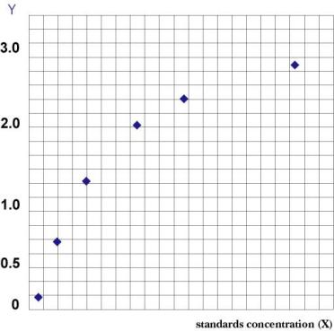
Representative Standard Curve
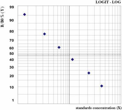
Detailed validation data (spike‑recovery, dilution linearity) are available in the product instruction manual.
Kit Components
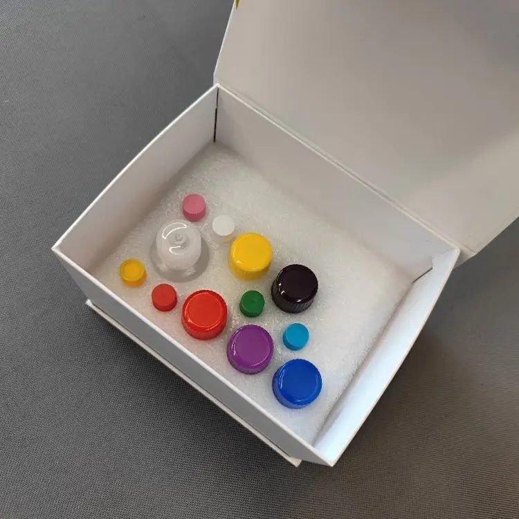
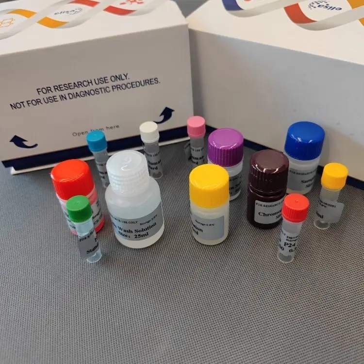
96‑Well Format
| Component | Quantity | Notes |
|---|---|---|
| Microelisa Stripplate | 12 strips × 8 wells | Pre‑coated with anti‑IP3 antibody |
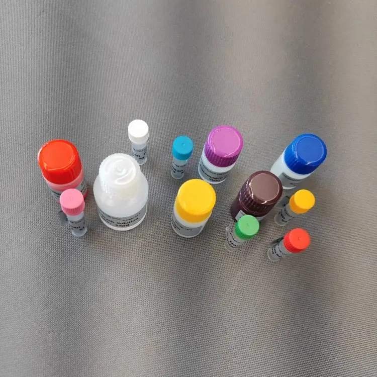
| Standard (0.3 mL/vial) | 6 vials | Ready‑to‑use liquid, 0–480 pg/mL |
| Sample Diluent | 6 mL | – |
| HRP‑Conjugated IP3 | 10 mL | Ready‑to‑use |
| 20× Wash Buffer | 25 mL | Dilute to 1× before use |
| Substrate A | 6 mL | – |
| Substrate B | 6 mL | – |
| Stop Solution | 6 mL | – |
| Plate Sealers | 2 sheets | – |
| Instruction Manual | 1 copy | – |
| Self‑sealing Bag | 1 piece | – |
48‑Well Format
| Component | Quantity | Notes |
|---|---|---|
| Microelisa Stripplate | 12 strips × 4 wells | Pre‑coated |
| Standard (0.3 mL/vial) | 6 vials | Ready‑to‑use |
| Sample Diluent | 3 mL | – |
| HRP‑Conjugated IP3 | 5 mL | Ready‑to‑use |
| 20× Wash Buffer | 15 mL | Dilute to 1× |
| Substrate A | 3 mL | – |
| Substrate B | 3 mL | – |
| Stop Solution | 3 mL | – |
| Plate Sealers | 2 sheets | – |
| Instruction Manual | 1 copy | – |
| Self‑sealing Bag | 1 piece | – |
Crystallization of 20× Wash Buffer upon cold storage is normal; warm in a water bath until crystals dissolve before dilution.
Assay Procedure Summary
- Equilibrate: Bring the foil pouch to room temperature for 20 min. Remove required strips.
- Add Standards & Samples: 50 μL standards; 10 μL sample + 40 μL Sample Diluent. Blank: leave empty.
- Add HRP‑IP3 Conjugate: 100 μL to all wells except blank. Incubate at 37°C for 60 min.
- Wash: Aspirate, wash with 1× Wash Buffer (soak 1 min), repeat 5 times.
- Substrate: 50 μL each of Substrate A and B, incubate dark at 37°C for 15 min.
- Stop & Read: 50 μL Stop Solution, read at 450 nm within 15 min.
Total hands‑on time: ~2 hours.
Throughput
| Format | Max. Samples (Single Wells) |
|---|---|
| 48‑well | 39–40 |
| 96‑well | 85–88 |
Related Products
Expand your extracellular matrix and cell signalling research with these complementary kits (available from Yanda Bio):
- Mouse 5-Hydroxytryptamine (5-HT) ELISA Kit – For Neuroscience & Neurotransmitter Research
- Human Syndecan‑4 (SDC‑4) ELISA Test Kit – For Cell Signaling & Wound Healing Researc
- Human CNP (C-type Natriuretic Peptide) ELISA Kit – the precise quantification of CNP in human serum, plasma, cell culture supernatants, and tissue homogenates.
- Human Hydroxymethylbilane Synthase (HMBS) ELISA Kit – This kit measures enzyme activity of human hydroxymethylbilane synthase (HMBS), also known as porphobilinogen deaminase (PBGD).
Frequently Asked Questions
Q: Is this a sandwich or competitive ELISA?
A: Competitive ELISA. IP3 is too small (~420 Da) for two antibodies to bind simultaneously. Free IP3 competes with HRP‑IP3 for antibody binding; signal is inversely proportional to concentration.
Q: Does this kit cross‑react with other inositol phosphates?
A: No significant cross‑reactivity with IP2, IP4, PIP2, or other analogues has been observed.
Q: How can I prevent IP3 degradation during extraction?
A: Add fresh phosphatase inhibitors (NaF 10–20 mM, orthovanadate 1 mM, or commercial cocktail). Process all samples on ice or at 4°C. Acid extraction with PCA/TCA followed by neutralization provides maximum stability.
Q: What is the recommended sampling time after cell stimulation?
A: IP3 peaks within seconds. Collect samples at 30 sec, 1 min, and 5 min post‑stimulation. Immediately quench with ice‑cold extraction buffer containing inhibitors.
Q: Can I use serum or plasma samples?
A: Not recommended. IP3 is an intracellular messenger and is rapidly degraded in blood. Cell lysates or tissue homogenates are the preferred sample types.
Q: Can this kit be used for mouse or rat IP3?
A: No. It is validated exclusively for human IP3. Species‑specific kits should be used for rodent samples.
Q: How do I calculate the final IP3 concentration?
A: Final concentration = In‑well interpolated value (from inverse standard curve) × 5 (in‑well dilution) × additional pre‑dilution factor.
Q: Are MSDS and composition documents available for export?
A: Yes. These documents are provided upon request and can be included with shipment.
Other FAQ For ELISA Test
Ordering & Contact
| Item | Details |
|---|---|
| Catalog Number | Yanda‑Human‑IP3 |
| Formats | 48‑well / 96‑well |
| Package Dimensions | 16 cm × 12.5 cm × 9.5 cm (neutral white box) |
| Shipping | Cold‑chain (2–8°C), protected from light |
| Storage | 2–8°C; do not freeze |
| Shelf Life | 6 months |
WhatsApp: +86 198 3186 0669 | Email: songhawyble@gmail.com
Technical Documents & Support
- Instruction Manual – Download or request via email.
- MSDS & Certificate of Composition – Available upon request.
- Technical Support – For inhibitor protocols, acid extraction, or stimulation time‑course design, contact us via WhatsApp or email.
Disclaimer
For research use only. Not for clinical diagnosis or treatment. Users are responsible for validating assay performance in their own experimental conditions. The manufacturer assumes no liability for improper use or protocol deviations.